


This article is the first in a two-part series.
Genetics is a fundamental field of biology that covers the passage of genetic information to successive generations (inheritance). More broadly, genetics covers the study of genes, how they are organized in cells, how they are regulated, and how they interact with each other and the environment. Genetics is a key component of genomics, a much broader area of the life sciences that is highly interdisciplinary and considers the structure, function, mapping, and both natural and induced alterations of genomes.
Over the course of a series of short articles, we will cover the basics of what DNA is and how it is organized in cells, how cells divide and pass genetic information to the next generation, the laws of inheritance, the molecular basis of disease, how purebred dogs can help advance genetic research, and how genetic variation (inherited and sporadic) impacts disease.
What is DNA and how is it organized in cells?
Cells are the basic structural and functional units of all living organisms. Inside all animal cells (with the exception of red blood cells) is a nucleus that contains the genetic material, deoxyribonucleic acid (DNA). A key component of DNA is the nucleotide. There are four nucleotides: adenine (A), thymine (T), guanine (G), and cytosine (C), and it is the specific order of these nucleotides that determines the genetic code.
- The genome of a dog contains approximately 2.4 billion nucleotides.
- If all the DNA inside one dog cell was laid out end to end, it would stretch over 6 feet in length.
- If all the DNA in the cells of one adult dog was laid out end to end, it would stretch to the sun and back many times.
A genome can be thought of as an instructional manual. The letters in the manual represent the nucleotides of the DNA, the words represent the genes, and the chapters represent the chromosomes. When the words are spelled correctly, and in the right order, they make sense and tell a genetic story.
To allow all the DNA from a single cell to fit inside a nucleus, a special protein scaffold allows the DNA to wind tightly into bundles. These bundles of protein and DNA, referred to as chromosomes, can then fit inside the nucleus. Chromosomes behave as nature’s biological filing cabinets, with animals of the same species having the same number of chromosomes in each cell and the same DNA within each chromosome.
In all animal cells (except egg and sperm cells), each chromosome is present twice, with one full set inherited from the mother and the other full set inherited from the father. Organisms with two copies of each chromosome are referred to as diploid. In reproductive cells (egg and sperm), each chromosome is present once. In all mammals, there are two chromosomes, X and Y, that determine the sex of an individual. Females have two copies of the X chromosome; males have one X and one Y chromosome. The rest of the chromosomes in a nucleus are called autosomes and are generally numbered, according to their size and shape, with the largest autosome called number 1, the second largest number 2, and so on.
The organization of the canine genome.
For many years, it has been known that the DNA of the canine genome is organized into 78 chromosomes per somatic (non-gamete) cell (39 in gametes; egg and sperm cells), present as 38 pairs of autosomes with either two copies of the X chromosome in females or one X and one Y in males [1]. Only males have the Y chromosome, and all female eggs have an X chromosome. This means that the sex of offspring is determined by whether the sperm that fertilizes the egg carries an X or a Y chromosome. From Affenpinscher to Yorkshire Terrier, all healthy dogs have 78 chromosomes, and under a microscope the chromosomes from each cell within a single dog and between dogs look the same. It is the actual DNA sequence where differences between breeds are detected.
Chromosomes can be stained with a dye that allows them to be seen under a specialized microscope when at a stage of cell division called metaphase (Figure 1). Specific dyes can be used to induce a banding pattern for each and then observed with a powerful microscope. Chromosomes of a dog cell are shown in Figure 2 below. Since each chromosome is present twice (maternal and paternal sets), the chromosome can be aligned into pairs, numbered accordingly, and displayed as shown in Figure 3.
Figure 1. Metaphase chromosomes of the domestic dog as they appear when stained with a blue fluorescent dye that allow them to be seen with a high-powered microscope. Each of the 78 rod-shaped structures are separate chromosomes. The two sex chromosomes (X and Y) are labeled, indicating that this cell is from a male.
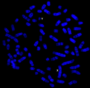
Figure 2. Metaphase chromosomes of the domestic dog when stained with a specific dye and viewed with a high-powered microscope to reveal banding patterns along the length of each chromosome. The banding patterns are the same for each pair, allowing the pairs to be identified. This is a male cell, as indicated by the presence of an X and Y chromosome (labeled).
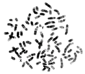
Figure 3. Chromosomes of the domestic dog from Figure 2 organized into 38 pairs, according to their banding pattern. In each pair, one is from the mother (egg) and one is from the father (sperm).
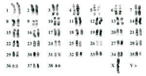
This arrangement of the chromosomes is referred to as a karyotype and serves as a common format for researchers and veterinary professionals to identify and refer to the individual chromosomes.
The precise order of the nucleotides that make up the canine genome sequence was determined in 2005, revealing a total of approximately 2.4 billion nucleotides [2]. The amount of DNA per chromosome was determined to range from approximately 126 million nucleotides for chromosome 1, to just 27 million for chromosome 38.
Learn more about genetics in part two.
Dr. Matthew Breen is a professor of Genomics and the Oscar J. Fletcher Distinguished Professor of Comparative Oncology Genetics in the Dept. of Molecular Biomedical Sciences at the NC State University College of Veterinary Medicine.
Cited publications.
1. Breen, M., J. Bullerdiek, and C.F. Langford, The DAPI banded karyotype of the domestic dog (Canis familiaris) generated using chromosome-specific paint probes. Chromosome Research, 1999. 7(5): p. 401-6.
2. Lindblad-Toh, K., et al., Genome sequence, comparative analysis and haplotype structure of the domestic dog. Nature, 2005. 438(7069): p. 803-19.