


This article is the second in a two-part series. Read part one.
Genetics is a fundamental field of biology that covers the passage of genetic information to successive generations (inheritance). More broadly, genetics covers the study of genes, how they are organized in cells, how they are regulated, and how they interact with each other and the environment. Genetics is a key component of genomics, a much broader area of the life sciences that is highly interdisciplinary and considers the structure, function, mapping and both natural and induced alterations of genomes.
Over the course of a series of short articles, we will cover the basics of what DNA is and how it is organized in cells; how cells divide and pass genetic information to the next generation; how genetic variation (inherited and sporadic) impacts disease; and how Here
How do cells divide?
Understanding the processes of cell division requires the definition of some key terms.
- Mitosis – Process of cell division of somatic cells in which a series of steps leads to the formation of two daughter cells that each has the same number of chromosomes as the parent cell
- Meiosis – Process of cell division with two stages, leading to the production of gametes in which the number of chromosomes is reduced to one half of the number in a somatic cell
- Haploid – Single set of chromosomes. In the dog, this is 38 autosomes plus one sex chromosome (either X in all females, or X or Y in males).
- Diploid – Double set of chromosomes. In the dog, this is 76 autosomes plus two sex chromosomes (either XX or XY).
- Somatic cell – Cell of a multicellular organism not associated with reproduction – (e.g. skin, bone, lung, liver, etc.)
- Gamete – Cell of a multicellular organism associated with reproduction (i.e. egg and sperm cells)
- Zygote – Cell formed by a fertilization event between an egg and sperm cell, combining the genetic material of these gametes.
From a single cell to the playful puppy that becomes part of our family and, for the rest of the dog’s life, the process of cell division is ongoing and carefully controlled. When a cell divides, it makes two cells (daughter cells), which then divide to make four cells, which divide to make eight cells, and so on. It is this exponential increase that leads to adult dogs comprising trillions of cells. During fetal development, the cells of a puppy are directed to form different types of cells, such a heart, brain, skeleton, kidney, lung, skin, nerve, blood, etc.
Once an adult, the extent of cell division alters, to repair injuries and replace dead cells. There is a very strict mechanism that signals when cells need to start and stop dividing. Different cell types divide at different rates. For example, millions of skin cells die each day and so the need to replace them means skin cells divide regularly. Other cells, such as nerve cells, divide much less frequently.
There are two types of cell division, referred to a mitosis and meiosis. Mitosis is the process by which somatic cells (non-reproductive) divide to replicate themselves. Meiosis is restricted to the germ cells (reproductive cells, egg and sperm) and is the process by which genetic variation is introduced into the next generation. The major difference between the processes of these alternate forms of cell division is the number of copies of each chromosome that the daughter cell each have. At the start of cell division, each cell has two copies of each chromosome and is referred to as diploid. In mitosis, each of the two daughter cells is diploid, whereas there are four daughter cells at the end of meiosis with each having half the number of chromosomes as the parent cell. These cells are referred to as haploid.
Mitosis.
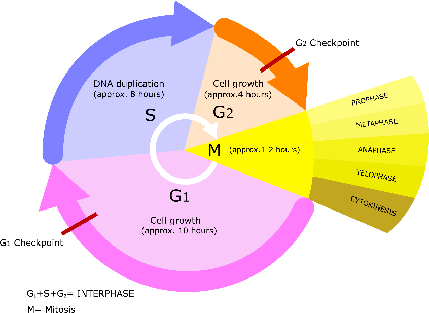
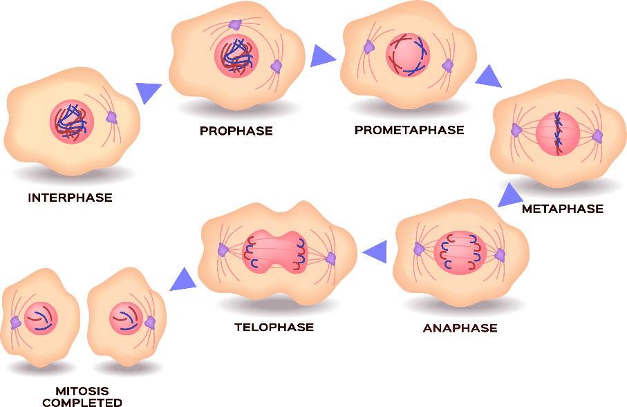
Each time a cell divides, it advances through the cell cycle (Figure 1), a process comprising a series of well-defined steps that need to happen for a cell to duplicate. While many somatic cells are generated daily, cells spend most of their time in interphase, where the cells gather all they need to divide, duplicate their DNA content to have sufficient to pass to each daughter cell. The actual process of mitosis (Figure 2) is a series of well-defined steps that each cell must take to divide into two daughter cells.
During the first three stages, prophase, prometaphase and metaphase, the chromosomes become progressively more condensed within the cell nucleus. By the end of metaphase, the chromosomes are aligned randomly along a structure called the metaphase plate, which spans the center of the nucleus. It is at the stage of metaphase that scientists trained at observing chromosomes will evaluate cells for changes to the expected number and structure of these elements. This is how many chromosomal aberrations are detected.
As the cell enters anaphase, special structures called spindle fibers have become attached to the centromeres of each chromosome. The fibers then pull apart the two halves (sister chromatids) of each chromosome, taking each half to opposite ends of the cell nucleus and the cell elongates. The chromosomes continue to condense during telophase and an enclosure (nuclear envelope) forms around the separated sets of chromosomes. The opposite ends of the cell continue to be pushed apart, and the cell then cleaves into two daughter cells.
Meiosis is the process of cell division that results in the gametes (egg and sperm). A major role of meiosis is to provide a mechanism, called genetic recombination, that facilitates the introduction of genetic variation to be passed to the next generation. The result of meiosis, the gametes, have only half the number of chromosome (a haploid set) of a somatic cell, and so there needs to be a fusion with another gamete of the same species to generate a new individual with a full complement of the genetic material for that species.
Meiosis is a process that occurs over two cycles, meiosis-I and meiosis-II (Figure 3). Before entering meiosis, the cell behaves much the same as in mitosis, duplicating its DNA content and gathering what it needs to divide. It is during meiosis-I that the process of genetic recombination occurs, providing opportunities for DNA from the two chromosomes of each pair to exchange information.
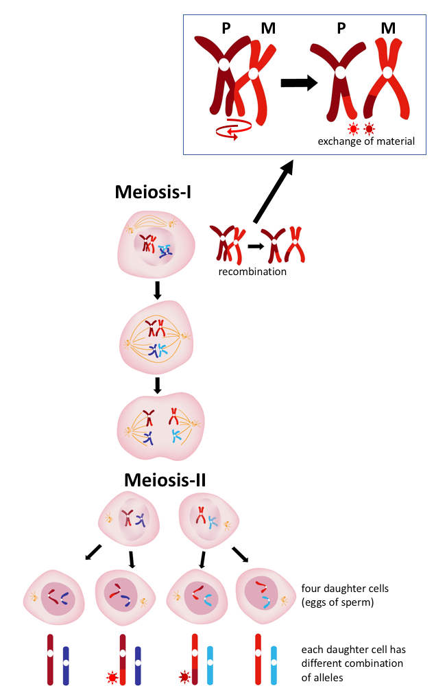
This process creates a new set of alleles for the genes along the length of each chromosome. Meiosis-II proceeds very much like mitosis. Since this happens in the development of both gametes, the number of possible combinations emerging from a fusion of sperm and egg is enormous and explains the vast breadth of phenotypes. Puppies from a litter may look and behave very much like each other initially, but as they grow and develop, we notice differences among them that allows us to recognize them for the unique genetic entities they are.
Dr. Matthew Breen is a Professor of Genomics and the Oscar J. Fletcher Distinguished Professor of Comparative Oncology Genetics in the Dept. of Molecular Biomedical Sciences at the NC State University College of Veterinary Medicine